Table of Contents
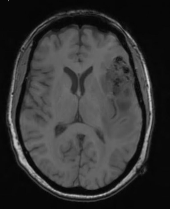
Washington University Experience | NEOPLASMS (GLIAL) | Glioblastoma, adenoid pattern | 13A1 Gliosarcoma, adenoid (Case 13) T1 - Copy
Case 13 History ---- The patient was a 59-year-old man who presented with aphasia. MRI revealed a 6.0 x 3.2 x 5.9 cm ring-enhancing lobulated mass with surrounding vasogenic edema in the left frontotemporoparietal region. ---- 13A1 MRI studies show a hypointense mass in this T1-weighted non-contrast image.

