Table of Contents
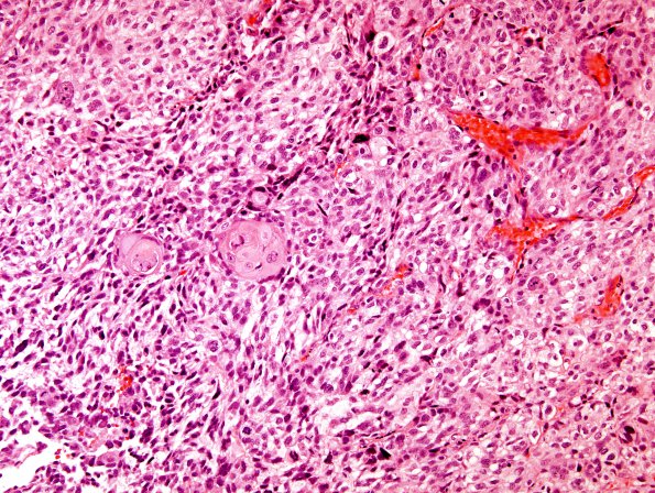
Washington University Experience | NEOPLASMS (GLIAL) | Glioblastoma, adenoid pattern | 14A1 GBM, adenoid features with pearls (Case 14) H&E 2
Case 14 History ---- The patient was a 63 year old man with a large cystic mass involving the skull and overlying scalp. Radiologically, the tumor extended to the adjacent dura, although there was no clear intracranial component. ---- 14A1,2 Sections reveal a highly cellular and markedly pleomorphic spindle cell neoplasm with small interspersed squamoid nests. The tumor cells display moderate quantities of clear to eosinophilic cytoplasm and contain oval to irregular nuclei with small nucleoli. The mitotic index is brisk. Focal basophilic material is also seen in some areas, suggestive of extra-cellular mucin. No definite tumor necrosis is noted, although the biopsy is relatively small.

