Table of Contents
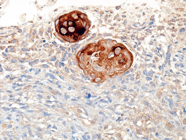
Washington University Experience | NEOPLASMS (GLIAL) | Glioblastoma, adenoid pattern | 14C2 GBM, adenoid features with pearls (Case 14) Cytomix
Stains for cytokeratin and epithelial membrane antigen (EMA) predominantly highlight the squamoid nodules. (EMA IHC) ---- Immunohistochemistry (not shown): The tumor cells are diffusely positive for vimentin. A stain for CAM 5.2 highlights the squamoid nests, although the majority of tumor cells were negative. The tumor was negative for smooth muscle actin, PSA, and PSAP. A histochemical stain for reticulin highlights dense intercellular deposition, consistent with a sarcomatous element. The morphologic and immunohistochemical features are consistent with the diagnosis of gliosarcoma with adenoid features; however, the lack of a clear intracranial component would make this diagnosis less likely. Therefore, the precise etiology of this tumor is unclear.

