Table of Contents
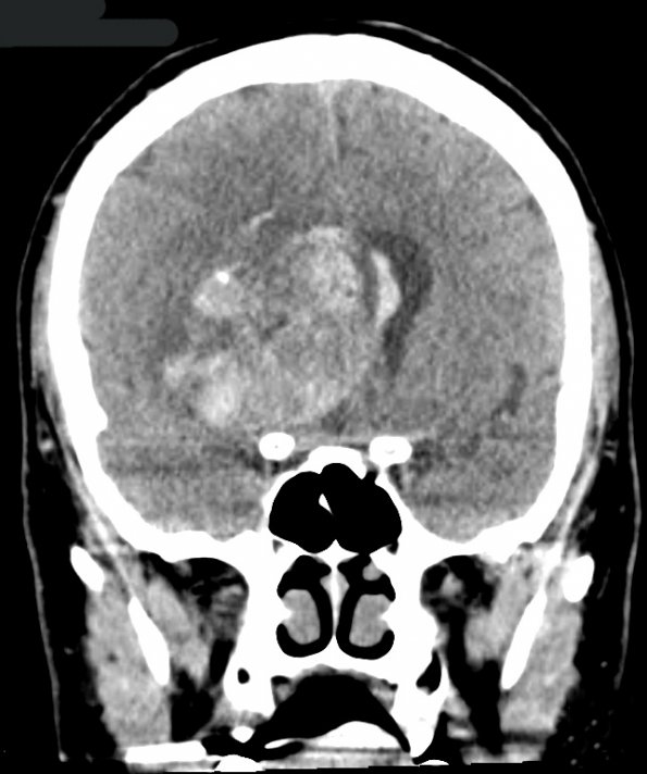
Washington University Experience | NEOPLASMS (GLIAL) | Glioblastoma, adenoid pattern | 15A1 Glioblastoma, adenoid type (Case 15) CT 1 - Copy
Case 15 History ---- The patient was a 47-year-old man who presented with complaints of fatigue for approximately 4-5 months. Approximately one month prior to admission, the patient noticed drooping of the left side of his mouth. MRI showed a predominantly ring-enhancing, centrally hemorrhagic mass centered in the right basal ganglia, measuring 4.5 x 4.3 x 3.3 cm with regional mass effect, subfalcine herniation, and left-to-right midline shift. Surrounding T2/FLAIR hyperintensity extended anteriorly into the deep white matter. Additionally, two small enhancing lesions within subcortical white matter of the right temporal lobe were separate from the main mass concerning for small satellite lesions or multifocality of the dominant lesion. Operative procedure: Right frontal stereotactic biopsy. ---- CT & MRI Studies: 15A1 CT showing hemorrhage, mass effect, and obliteration of the right ventricle.

