Table of Contents
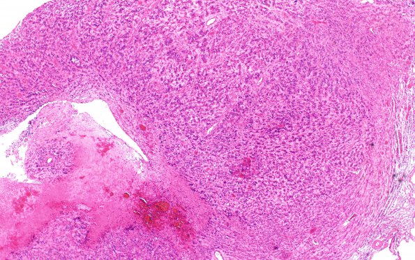
Washington University Experience | NEOPLASMS (GLIAL) | Glioblastoma, adenoid pattern | 2A1 Glioblastoma, adenoid (Case 2) H&E 4X
Case 2 History ---- The patient was a 38 year old woman originally evaluated for a left hemispheric mass which was excised and a diagnosis of glioblastoma was made. She later presented with a right posterior chest wall mass suspected to represent metastatic malignancy. ---- 2A1-4 The sections of the left hemispheric brain mass and right pleural mass both show a hypercellular neoplasm with extensive areas of geographic necrosis. At the transition of viable tumor and necrosis, there is prominent endothelial vascular proliferation. The tumor cells are arranged in an intertwining to trabecular pattern. In some regions, the tumor cells are radially aligned around individual vascular structures simulating pseudorosettes. Some tumor cells show slender, spindle-shaped nuclei suggestive of a sarcomatous component. A majority of the tumor cells, however, are epithelioid and contain round to oval, markedly pleomorphic nuclei. Mitotic activity is abundant.

