Table of Contents
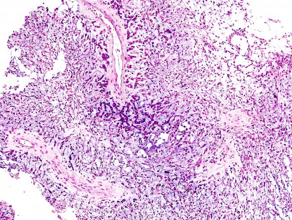
Washington University Experience | NEOPLASMS (GLIAL) | Glioblastoma, adenoid pattern | 3A1 GBM, Adenoid (Case 3) H&E 7
Case 3 History ---- The patient was a 63 year old man with history of progressive left facial weakness, gait imbalance and headache for approximately 10 days. CT demonstrated a cystic 5.4 cm right parietal mass with an irregular margin and vasogenic edema. MRI showed a rim enhancing mass of the right temporal lobe measuring 6.3 x 4.8 x 4.4 cm, with a ~2 x 2.8 cm nodular enhancing portion laterally, and surrounding edema that extended into the basal ganglia, the Sylvian area, and the posterior right periventricular white matter. Operative procedure: Right temporal craniotomy for resection of right temporal parietal brain tumor. ---- 3A1,2 The majority of H&E stained sections show a high grade cellular neoplasm with classic 'glioblastoma-like' features, including gemistocytes, atypical hyperchromatic 'naked' nuclei with indiscernible cytoplasm, abundant mitotic figures, microvascular proliferation / endothelial hyperplasia, thrombosis, and areas of necrosis (geographic and pseudopalisading). However, a sizable component of the sampled tissue shows two other unusual patterns which exhibit a pale, basophilic myxoid material in which eosinophilic tumor cells appear singly, in cords, and in clusters. In many areas, this pattern is reminiscent of chordoma, chordoid meningioma, or mucinous ('colloid') carcinoma. Within the context of glioblastoma, this pattern suggests a diagnosis of glioblastoma with adenoid features. According to current data, this distinction does not appear to alter prognosis. (H&E)

