Table of Contents
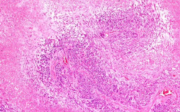
Washington University Experience | NEOPLASMS (GLIAL) | Glioblastoma, adenoid pattern | 4A3 Glioblastoma, adenoid (Case 4) H&E 10X
Sections of the mass showed a glioblastoma multiforme with the histologic features of a glioblastoma, adenoid pattern. In these areas there is extensive necrosis within which are perivascular islands of hyperchromatic, epithelioid cells that radiate out from the vasculature. In foci these cells form sheets and nests. In the necrotic areas ghosts of neoplastic cells can be seen. There are also areas of myxoid appearing degeneration. In other areas the tumor has the typical appearance of an astrocytic neoplasm with marked hypercellularity, pleomorphism, vascular proliferation and diffuse infiltration of the surrounding brain parenchyma.

