Table of Contents
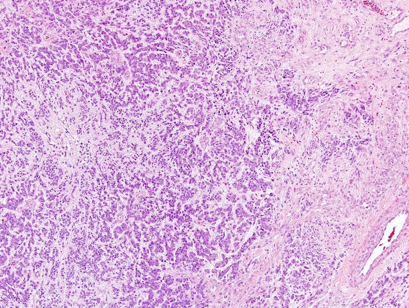
Washington University Experience | NEOPLASMS (GLIAL) | Glioblastoma, adenoid pattern | 7A1 GBM, Adenoid Features (Case 7) H&E 3
Case 7 History ---- The patient was a 75 year old man who developed forgetfulness and confusion after a febrile episode; however, he continued to carry on with his activities of daily living without significant disturbance. An MRI and CT showed a large (5 x 4 x 4 cm) cystic mass at the left parietal-occipital junction with significant mass effect on the left cerebral peduncle and surrounding brain. Operative procedure: Craniotomy for tumor. ---- 7A1-3 This glioblastoma presents a complicated and unusual histologic picture. Most of the tumor consisted of undifferentiated cells with scant cytoplasm and small oval to angulated nuclei with a granular, evenly dispersed chromatin pattern. In many areas, the tumor was quite organized in its architecture with nests and ribbons of cells separated by a myxoid stroma. This imparts an adenoid appearance to the tumor. Recognition of this tumor as GBM rather than a metastatic tumor was aided by the presence of areas consisting of cells with more abundant cytoplasm and disorderly arrangement typical of GBM, i.e., with vascular proliferation and areas of necrosis.

