Table of Contents
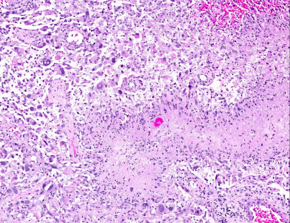
Washington University Experience | NEOPLASMS (GLIAL) | Glioblastoma, adenoid pattern | 8A6 GBM, adenoid (epithelioid) form (AANP 1990, Case 6) H&E X10 4
In some areas there is pseudopalisading necrosis and small undifferentiated cells forming a histopathologic appearance of more classical GBM. (H&E) ---- Focal areas in the tumor stained positively for reticulin. The tumor was strongly GFAP positive and cytokeratin negative. The diagnosis was adenoid glioblastoma multiforme.

