Table of Contents
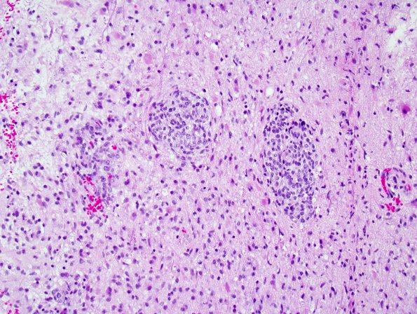
Washington University Experience | NEOPLASMS (GLIAL) | Glioblastoma, adenoid pattern | 9B1 GBM, Adenoid (Case 9) H&E 1.jpg
9B1,2 These images show several small epithelioid nests resembling metastatic carcinoma. Clusters consisted of enlarged cells with more definitive cell borders and moderate degrees of anaplasia. Some of these are distributed in a perivascular fashion, whereas others appear to be within the parenchyma. Surrounding the nodules are fragments of brain parenchyma involved by a moderately cellular infiltrative glial neoplasm. There is moderate nuclear pleomorphism, with the majority of tumor cells resembling fibrillary and gemistocytic astrocytoma cells, including elongate, irregular, and hyperchromatic nuclei with variable quantities of eosinophilic cytoplasm. Scattered mitotic figures are seen and there are small foci of incipient microvascular proliferation and necrosis.

