Table of Contents
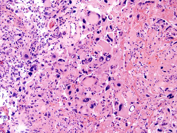
Washington University Experience | NEOPLASMS (GLIAL) | Glioblastoma, giant cell | 1A1 GBM, Giant Cell Type (Case 1) H&E 4
Case 1 History ---- The patient was a 30 year old man with a 3.8 x 2.7 x 2.8 cm focally rim-enhancing left frontal lobe lesion, who received steroids at an outside institution. Operative procedure: Craniotomy and biopsy. ---- 1A1,2 The neurosurgical specimen consists of numerous multinucleated bizarre giant cells with variably sized hyperchromatic nuclei and eosinophilic cytoplasm admixed with small cells with bipolar nuclei. There are multifocal areas of necrosis with pseudo-palisading of tumor cells but not endothelial proliferation.

