Table of Contents
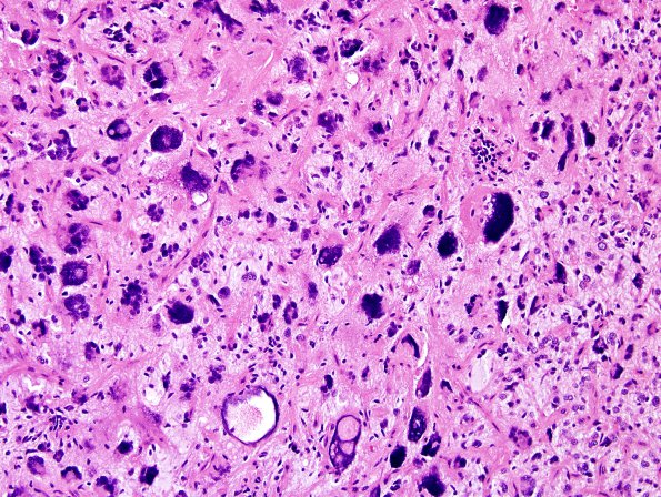
Washington University Experience | NEOPLASMS (GLIAL) | Glioblastoma, giant cell | 2A1 GBM w Giant Cell Features (Case 2) H&E 5.jpg
Case 2 History ---- The patient was a 68-year-old woman eight years status-post right partial mastectomy for ductal carcinoma in situ and one year status-post left partial mastectomy for ductal carcinoma in situ arising within a papilloma. Following her right surgical therapy, she completed brachytherapy with partial breast radiation and completed adjuvant with partial breast radiation. ---- Two years later she presented with a “cerebrovascular accident”. Four weeks after that she developed increased weakness of her left leg and the right side of her face was numb. Imaging workup suggested her CVA was, in actuality, a brain tumor which was biopsied. ---- 2A1-3 The tumor consists of a high-grade, infiltrating glial neoplasm characterized by large numbers of small, oval, astrocytic-line elements as well as a second complement of a large number of multinucleate/giant cells. There was endothelial proliferation and a few foci of necrosis with pseudo-palisading. Numerous mitoses are seen, involving both small and giant cells.

