Table of Contents
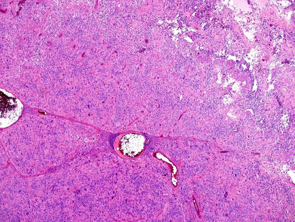
Washington University Experience | NEOPLASMS (GLIAL) | Glioblastoma, giant cell | 6A1 GBM, giant cell and PNET (Case 6) H&E 4X.jpg
Case 6 History ---- The patient was a 46 year old woman. No additional clinical history was provided or present in the available medical records. ---- 6A1-4 Sections of the resected right frontal brain tumor show a diffuse glioma with pseudopalisading necrosis and microvascular proliferation. Neoplastic cells basically fall into several distinct categories. The first population is made up of markedly enlarged, highly atypical, frequently multinucleated tumor cells with abundant glassy eosinophilic cytoplasm. A second population appears in hypercellular clusters of mitotically active tumor cells with high nucleus to cytoplasm ratio and comparatively mild pleomorphism. This second population represents glioblastoma with a primitive neuronal component (see section in this atlas for the presentation of additional material from this case). Nuclei from both groups exhibit hyperchromasia with finely granular evenly dispersed chromatin and multiple inconspicuous nucleoli. Additional smaller cells have hyperchromatic nuclei, some bipolar.

