Table of Contents
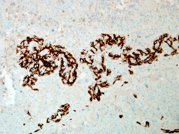
Washington University Experience | NEOPLASMS (GLIAL) | Glioblastoma, giant cell | 6G4 GBM, giant cell and PNET (Case 6) CD34 endo prolif 20X.jpg
Higher magnification of proliferated vessels. (CD 34 IHC). ---- Additional immunohistochemistry and FISH results: Synaptophysin is positive throughout the tumor, but the signal is stronger in areas resembling primitive neurons. Immunohistochemical stain for mutant IDH-1 (p.R132H) is negative. Immunohistochemical stain for mutant BRAF (p.V600E) was negative. FISH studies showed no evidence of EGFR gene amplification, although there was polysomy of chromosome 7 in 45% of cells. Assessment of loss of 10q (PTEN) was not informative. There was no co-deletion of the chromosomal regions 1p and 19q. There was no amplification of the N-MYC or C-MYC genes. ---- The morphology of this tumor strongly suggests a giant cell component. In agreement with this impression, there was widespread p53 immunoreactivity. A second population described above has histomorphologic and immunohistochemical signs of a primitive neuronal component and is presented further in glioblastoma, with primitive neuronal section of the atlas.

