Table of Contents
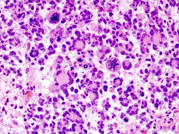
Washington University Experience | NEOPLASMS (GLIAL) | Glioblastoma, giant cell | 7A1 GBM, Giant cell type (Case 7) H&E 3
Case 7 History ---- The patient was a 26-year-old man with no known past medical history who developed a new-onset tonic-clonic seizures leading to an automobile accident. Subsequent imaging at an outside institution revealed a lesion in the left frontal lobe, suspicious for neoplasm. Magnetic resonance imaging at BJH showed a 2.9 x 3.0 x 3.2 cm heterogeneously enhancing intra-axial left frontal mass with surrounding vasogenic edema, and no central restricted diffusion to suggest an abscess. Clinical diagnosis: High grade neoplasm. Operative procedure: Left frontal craniotomy with resection. ---- 7A1,2 The tumor cells comprising the lesion have several morphologies. Most striking are abundant multinucleated touton-type giant cells with a 'wreath' of eccentrically located nuclei surrounding centrally located cytoplasm. Occasionally, these cells have bizarre highly anaplastic nuclei. Other cells have features of gemistocytes, with abundant eosinophilic opalescent cytoplasm and eccentrically located reniform or pleomorphic nuclei. Scattered amongst these large tumor cells are smaller bipolar tumor cells with elongated nuclei and thin cytoplasmic processes. The quality of the background parenchyma appears variably myxoid, and some areas show microcystic pools of mucin. Mitoses are easily identified. Endothelial hyperplasia is widespread. The tumor has focal areas of dyscohesion that are suggestive of necrosis, but no definitive necrosis is identified.

