Table of Contents
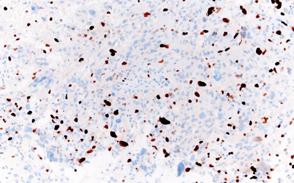
Washington University Experience | NEOPLASMS (GLIAL) | Glioblastoma, giant cell | 9E2 Giant Cell GBM (NP18-609) Ki67 20X
Higher magnification image (GFAP IHC) ---- Immunohistochemical stains (not shown): P53 and IDH1 (R132H) are negative in the tumor cells. The tumor cells are also negative for H3K27M and have retained H3K27me3 expression. Ki-67 proliferative index is estimated to be ~10 to 20%. There is no increase in reticulin deposition. ---- FISH studies show 10q loss with no evidence of EGFR amplification. MGMT promoter methylation is absent. Foundation One molecular profiling demonstrates mutations in TSC1 and a P53 splice site but no changes in EGFR, PDGFRA or IDH1. These histomorphological, immunohistochemical, and cytogenetic findings support the "integrated" diagnosis: giant cell glioblastoma, IDH1 (p. R132H) negative by immunohistochemistry, WHO grade 4.

