Table of Contents
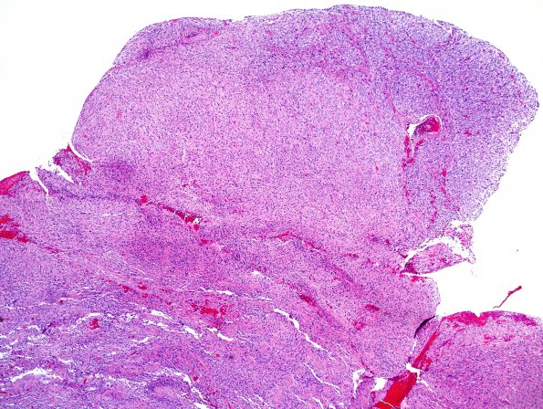
Washington University Experience | NEOPLASMS (GLIAL) | Glioblastoma, primitive neuronal component | 10A1 GBM, focal PNET (Case 10) H&E 4X.jpg
Case 10 History ---- The patient is a 13 year old boy. ---- Sections from the intraventricular tumor show numerous small tumor cells with cytological atypia, pleomorphic nuclei (often with bipolar nuclei), pseudopalisading necrosis, endothelial proliferation and prominent mitotic activity. In addition, other areas show dense cellularity with a high nuclear:cytoplasmic ratio and primitive neuroectodermal tumor-like morphology. ---- 10A1 Whole mount of the tumor shows foci of dense cellularity at the superior aspect of the image (H&E)

