Table of Contents
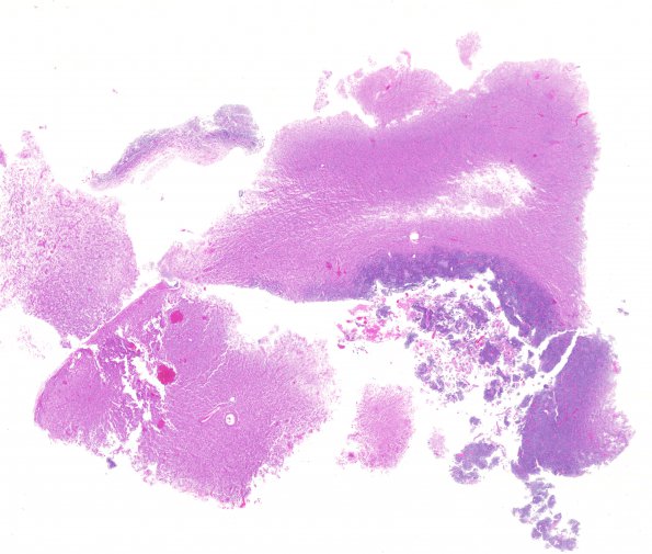
Washington University Experience | NEOPLASMS (GLIAL) | Glioblastoma, primitive neuronal component | 12A1 GBM, PNET (Case 12) 1 H&E WM
Case 12 History ---- The patient was a 59 year old man who presented with drowsiness and hemiparesis of 4-5 days' duration and was found to have a right frontoparietal mass with associated hemorrhage on imaging. Operative procedure: Resection. ---- 12A1 Sections reveal a moderate to highly cellular neoplasm with two distinct morphologic appearances. In some areas, the tumor appears infiltrative and has features consistent with a fibrillary and gemistocytic astrocytoma. There is no definite microvascular proliferation or necrosis in this area. ---- However, there are also relatively discreet nodules that display markedly increased cellularity and are composed predominantly of small primitive appearing cells. This component displays a high mitotic index with numerous pyknotic nuclei. Cell molding is prominent in these areas. Additionally, portions of this component grow in a nodular pattern with focal formation of delicate fibrillary processes, resembling neuropil. Vague structures resembling Homer Wright rosettes are also evident. Microvascular proliferation and tumor necrosis are both seen in this component.

