Table of Contents
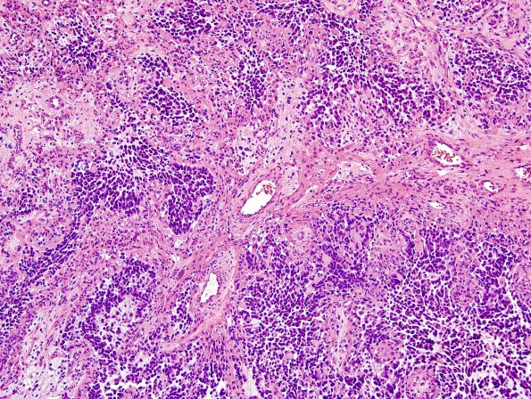
Washington University Experience | NEOPLASMS (GLIAL) | Glioblastoma, primitive neuronal component | 14A1 GBM, (Case 14) H&E 10X 1
Case 14 History ---- The patient was a 62-year-old man with essential tremor who developed several weeks of intermittent facial droop and headache. MRI showed a 6.4 cm solid and cystic right temporal lobe mass with surrounding vasogenic edema, midline shift and subfalcine and uncal herniation. Operative procedure: IMRI right craniotomy for tumor. ---- 14A1,2 This was malignant glioma with extensive geographic and pseudopalisading necrosis and extensive microvascular proliferation around fibrovascular cores ---- . Epithelioid tumor cells grow along and are separated by broad mesenchymal cores made up of spindle cells, some set within a myxoid background. Admixed with epithelioid cells are primitive cells with high N/C ratio and very frequent mitoses.

