Table of Contents
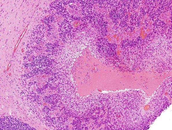
Washington University Experience | NEOPLASMS (GLIAL) | Glioblastoma, primitive neuronal component | 16A1 GBM, with PNET features (Case 16) H&E 5
Case 16 History ---- The patient was a 61 year old man presenting with left-sided numbness and weakness. MRI showed a 4.4 x 3.4 x 4.4 cm rim-enhancing cystic mass with a heterogeneous nodule in the posterior right frontal lobe. Operative procedure: craniotomy and resection. ---- 16A1-3 This is a high grade glial neoplasm composed predominantly of primitive appearing cells. Overall, the primitive areas appear solid and well-circumscribed, but a lower grade more infiltrative component is seen at the periphery of the mass. The infiltrative component appears more glial with a pleomorphic population of astrocytic cells. Microvascular proliferation and pseudopalisading necrosis are also present. The majority of the PNC cells are large with minimal cytoplasm, numerous mitotic figures, and apoptotic nuclei.

