Table of Contents
Washington University Experience | NEOPLASMS (GLIAL) | Glioblastoma, primitive neuronal component | 1A1 GBM, focal PNET (Case 1) H&E WM copy.jpg
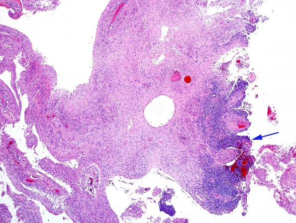
Case 1 History ---- The patient was a 59 year old woman who presented with altered mental status and confusion. Head CT and brain MRI showed a 6 cm cystic and solid mass predominating in the right frontal lobe, extending across the midline into the left frontal lobe, resulting in right-to-left subfalcine herniation, bilateral mesiotemporal lobe downward herniation and obstruction of the outflow of the left lateral ventricle at the level of the foramen of Monro. Operative procedure: Excision. ---- H&E sections of the right frontal lobe resection material show a complex high grade glial neoplasm with several distinct diagnostically-relevant histological patterns. The dominant histological pattern is that of glioblastoma with focal collections of primitive elements. ---- 1A1 A whole mount of the neurosurgical specimen showing diffuse glioblastoma with focal primitive neuronal collections (arrow) (H&E)

