Table of Contents
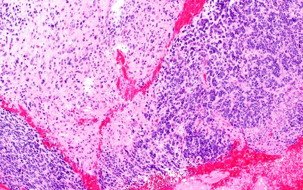
Washington University Experience | NEOPLASMS (GLIAL) | Glioblastoma, primitive neuronal component | 20A1 GBM w PNET Diffn (Case 20) H&E 1
Case 20 History ---- The patient was a 73 year old man with a ring-enhancing lesion of the temporal lobe. Operative procedure: Stereotactic guided left craniotomy for tumor resection. ---- 20A1 Areas of glioblastoma and PNC are seen in the specimen. (H&E). There is a population of markedly atypical cells with irregular pleomorphic nuclei, multi-nucleated cells, focal necrosis, and vascular proliferation. These features are consistent with a glioblastoma. Focally, the lesion also has some nodules composed of smaller cells with minimal cytoplasm, which are consistent with primitive neuronal cells.

