Table of Contents
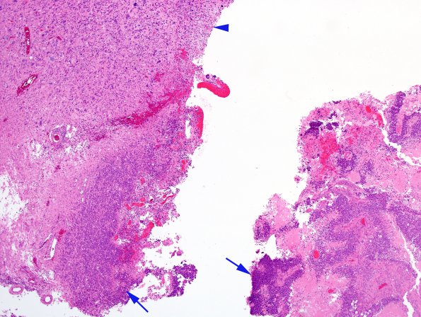
Washington University Experience | NEOPLASMS (GLIAL) | Glioblastoma, primitive neuronal component | 2A1 GBM, PNET (Case 2) Junction A 4X H&E copy.jpg
Case 2 History ---- The patient was a 51-year-old man who presented to an outside hospital with severe headaches after experiencing one month of balance and concentration difficulties. Multiple CT and MRI studies showed a large mass with heterogeneous enhancement involving the right lateral ventricle and right paraventricular white matter. Operative procedure: Right parietal craniotomy for tumor with intraoperative MRI. ---- 2A1,2 H&E stained sections show a high grade neoplasm with several dominant histological patterns. The first pattern (arrowhead, 2A1) is consistent with high grade glioma and features sheets of densely packed spindled tumor cells with endothelial hyperplasia and pseudopalisading necrosis. ---- The second pattern (arrows, 2A1) is morphologically most consistent with a primitive neuronal complement. These tumor cells have minimal cytoplasm and hyperchromatic stippled nuclei. (H&E)

