Table of Contents
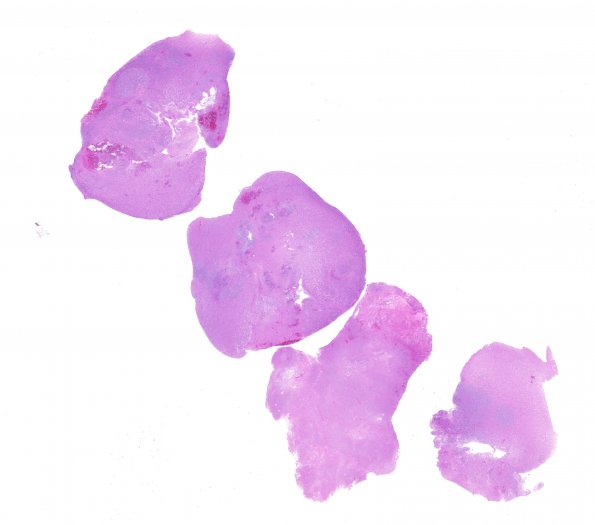
Washington University Experience | NEOPLASMS (GLIAL) | Glioblastoma, primitive neuronal component | 4A1 GBM with PNET features (Case 4) 1 H&E whole moun
Case 4 History ---- The patient was a 61-year-old woman who developed a headache related to a motor vehicle accident; computed tomography (CT) without contrast showed subarachnoid hemorrhage. Her headaches persisted, and repeat head CT without contrast showed a new hypodense mass in the medial right frontal lobe. Follow up MRI showed a 5.7 x 3.3 x 4.3 cm centrally necrotic mass with a thick irregular rim of enhancement in the right frontal lobe, also involving the right frontal horn of the lateral ventricle and crossing the genu of the corpus callosum. Operative procedure: Craniotomy for right frontal tumor excision. ---- 4A1,2 H&E stained sections show a diffuse high grade glioma of heterogeneous composition. Much of the viable tumor is formed by sheets of large highly pleomorphic cells with irregular, hyperchromatic nuclei; some with one nucleus and minimal cytoplasm, and others with one or more nuclei and abundant glassy eosinophilic cytoplasm. Mitotic figures in these areas are numerous, with several commonly appearing in a single 40x objective field. Microvascular proliferation, endothelial hyperplasia, and necrosis (both pseudopalisading and geographic) are readily identified. This histomorphologic pattern is characteristic of glioblastoma (GBM).

