Table of Contents
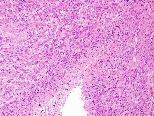
Washington University Experience | NEOPLASMS (GLIAL) | Glioblastoma, primitive neuronal component | 4A2 GBM with PNET features (Case 4) H&E 7.jpg
Much of the viable tumor is formed by sheets of large highly pleomorphic cells with irregular, hyperchromatic nuclei; some with one nucleus and minimal cytoplasm, and others with one or more nuclei and abundant glassy eosinophilic cytoplasm. Mitotic figures in these areas are numerous, with several commonly appearing in a single 40x objective field. Microvascular proliferation, endothelial hyperplasia, and necrosis (both pseudopalisading and geographic) are readily identified.

