Table of Contents
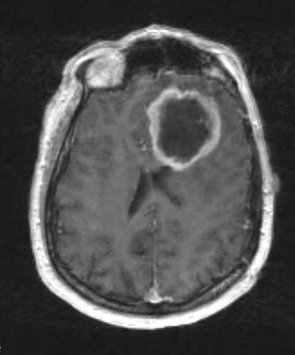
Washington University Experience | NEOPLASMS (GLIAL) | Glioblastoma, primitive neuronal component | 6A1 GBM w PNET features (Case 6) T1 W - Copy
Case 6 History ---- The patient was a 49 year old man with a past medical history of a diffuse astrocytic neoplasm WHO Grade 2 of the left temporal lobe. He was subsequently treated with radiation therapy and Temodar chemotherapy. Per clinical records he had increased confusion and speech difficulty after 7 years. MRI of the brain at that time showed a large well-defined T1 hypointense, T2 hyperintense mass in the inferior left frontal lobe with thick nodular peripheral ring enhancement. Operative procedure: Left frontal craniotomy with tumor resection. ---- 6A1 A T1-weighted contrast administered scan shows a large ring-enhancing mass which was biopsied.

