Table of Contents
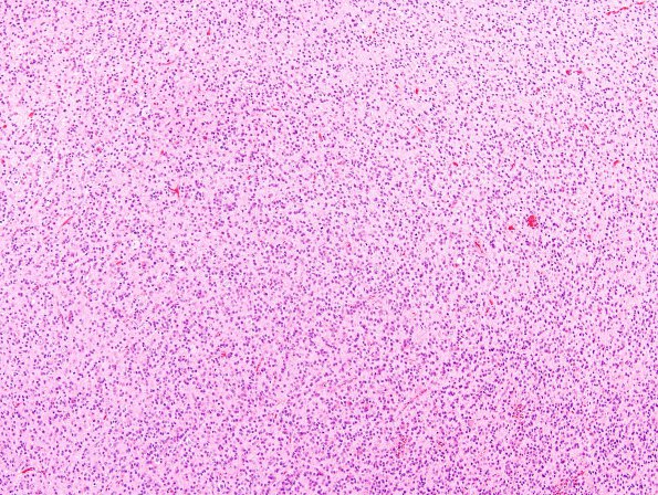
Washington University Experience | NEOPLASMS (GLIAL) | Glioblastoma, small cell type | 13A1 GBM, small cell (Case 13) H&E 9.jpg
Case 13 History ---- The patient was a 64 year old woman who presented with an inhomogenously enhancing left occipital lobe mass. ---- 13A1-4 This is an infiltrative glial neoplasm with marked cellularity and exuberant endothelial hyperplasia. The majority of the tumor cells have an oval nuclear contour with mild hyperchromasia and occasional perinuclear clear halos. Additionally, spindled nuclei and giant cells are seen. Some tumor cells have discernable eosinophilic cytoplasm. The mitotic index is brisk and there is focal necrosis. In some areas, there is a branching capillary network reminiscent of that seen in oligodendroglial neoplasms.

