Table of Contents
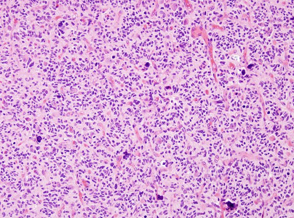
Washington University Experience | NEOPLASMS (GLIAL) | Glioblastoma, small cell type | 14A1 Glioblastoma, Small Cell (Case 14) H&E 8
Case 14 History ---- The patient was a 60 year old woman who presented with headache and 4.9 x 2.2 cm enhancing heterogenous left temporal lobe and epidural mass. Operative procedure: Left craniotomy and resection. ---- 14A1,2 This is a markedly cellular infiltrative glial neoplasm. Calcifications are prevalent, and there are areas with delicate, geometrically branching capillary networks. There is nuclear pleomorphism, with the majority of tumor nuclei appearing oval with mild hyperchromasia and minimal discernible cytoplasm. Despite the bland cytologic features, the mitotic index is markedly elevated. Endothelial proliferation is present. Necrosis is identified, but the pseudopalisading architecture is equivocal. The morphologic features are consistent with the small cell variant of glioblastoma, WHO grade IV.

