Table of Contents
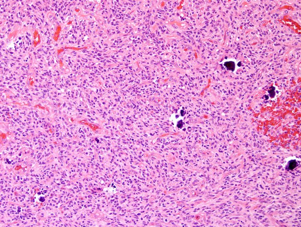
Washington University Experience | NEOPLASMS (GLIAL) | Glioblastoma, small cell type | 17A1 GBM, small cell type (Case 17) H&E 1.jpg
Case 17 History ---- The patient was a 72-year-old man with a brain tumor. MRI showed a 6.9 cm heterogeneously-enhancing solid and cystic mass in the posterior right temporal and parietal lobes with associated mass effect. Operative procedure: Right parietotemporal craniotomy for tumor. ---- 17A1-3 Neoplastic cells are arranged predominantly in hypercellular sheets with geographic necrosis and pseudopalisading necrosis. Glomeruloid microvascular proliferation is extensive and there is fibrinoid vascular necrosis. Neoplastic nuclei are relatively small with fairly uniform, round to oval nuclei, and variable degrees of hyperchromasia. The mitotic rate is very high, with up to six mitotic figures in a single high power field.

