Table of Contents
Washington University Experience | NEOPLASMS (GLIAL) | Glioblastoma, small cell type | 18A1 GBM, small cell type (Case 18) TIRM 1
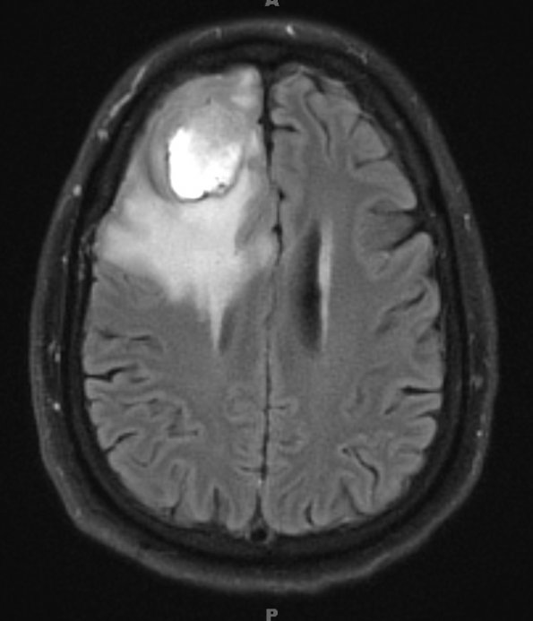
Case 18 History ---- The patient was a 55-year-old man who experienced new onset of seizures. MRI showed a 3.9 cm lesion in the right frontal lobe with peripheral nodular enhancement, central necrosis, and areas of increased perfusion. The principal radiographic differential diagnostic considerations included primary glial neoplasm versus metastasis. Operative procedure: Right frontal craniotomy for tumor. ---- 18A1-3 MRI examination is shown for FLAIR (18A1), T1-weighted contrast administered (18A2) and T2-weighted contrast administered (18A3) scans.

