Table of Contents
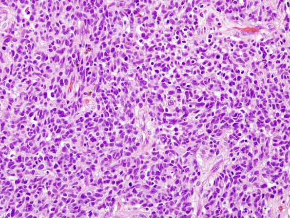
Washington University Experience | NEOPLASMS (GLIAL) | Glioblastoma, small cell type | 19A1 GBM, small cell variant (Case 19) H&E 1.jpg
Case 19 History ---- The patient was a 76-year-old man with an intra-axial, peripherally enhancing, hemorrhagic mass with nodular components centered at the right temporal lobe with extension into the parietal lobe, measuring 4.9 x 4 cm in size. There is surrounding T2/FLAIR hyperintense signal, indicating edema, as well as associated mass effect on the atrium and occipital horn of the right lateral ventricle. Operative procedure: Right craniotomy for resection of tumor. ---- 19A1-3 This is a hypercellular neoplasm composed of small cells with minimal cytoplasm, oval nuclei, and relatively fine chromatin with occasional small nucleoli. The nuclei show a mild amount of pleomorphism, with round to ovoid shape and mildly irregular contours. Scattered gemistocytic forms are also identified. On high magnification examination, mitotic figures are numerous, ranging up to 10 forms in a single HPF (40x). Additionally, this tumor shows endothelial proliferation and scattered examples of vascular fibrinoid necrosis. Frank areas of tumor necrosis are identified.

