Table of Contents
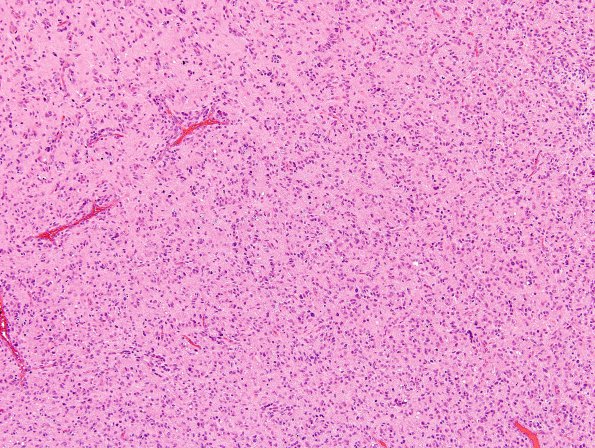
Washington University Experience | NEOPLASMS (GLIAL) | Glioblastoma, small cell type | 21A1 GBM, small cell variant (Case 21) H&E 9.jpg
Case 21 History ---- The patient was a 42 year old woman with a left frontal lobe neoplasm. A small cell GBM was favored on the resection by the originating institution specimen, though the possibility of an oligodendroglial component was considered. ---- 21A1-3 Sections revealed a highly cellular, infiltrative glial neoplasm characterized by large zones of necrosis as well as smaller foci of necrosis associated with nuclear pseudo-palisading. There is extensive endothelial hyperplasia and mitoses are seen in abundance. In regions of cortical involvement, there are foci of secondary structures, with perineuronal satellitosis and perivascular aggregation. The neoplastic cells are moderately pleomorphic, composed of predominantly oval nuclei with hyperchromasia and minimal cytoplasm. Occasional multinucleated cells and atypical mitotic figures are seen as well. Although some of the tumor cells harbored "rounder" nuclei, a definite oligodendroglial component was not found.

