Table of Contents
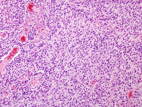
Washington University Experience | NEOPLASMS (GLIAL) | Glioblastoma, small cell type | 2A1 GBM , small cell features (Case 2) H&E 1.jpg
Case 2 History ---- This patient was a 50-year-old woman with a tumor in the occipital lobe. Histological differential diagnosis included anaplastic oligodendroglioma vs. small cell glioblastoma. ---- 2A1-3 Histological sections of the occipital brain tumor show a hypercellular glial neoplasm with widespread endothelial hyperplasia and pseudopalisading necrosis. Mitotic figures are abundant, often appearing at a density of four or more per high power field (HPF, 40X objective). The tumor cells are small, somewhat elongate, and relatively uniform, with a high nuclear-to-cytoplasmic ratio, mildly irregular nuclei, inconspicuous nucleoli, and scant eosinophilic cytoplasm. The stromal background appears coarsely vesiculated.

