Table of Contents
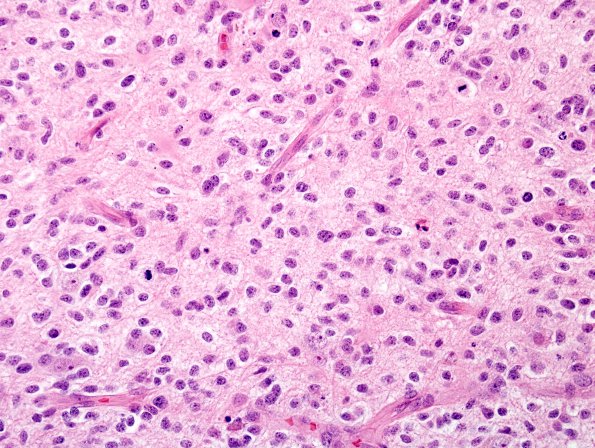
Washington University Experience | NEOPLASMS (GLIAL) | Glioblastoma, small cell type | 3A2 GBM , small cell features (Case 3) H&E 3.jpg
The tumor cells are oval with mild hyperchromasia and minimal discernible cytoplasm. The mitotic index is markedly elevated and there is extensive endothelial hyperplasia. ---- Ancillary studies (not shown): A special stain for GFAP immunoreactivity reveals fibrillary processes in a subset of tumor cells.
FISH studies showed normal dosages of chromosomes 1 and 19, i.e., no deletions were identified. There was evidence for gain of chromosome 7 in the absence of EGFR gene amplification. Although not absolutely specific, this pattern is consistent with that of glioblastoma.

