Table of Contents
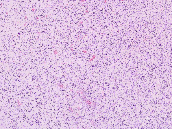
Washington University Experience | NEOPLASMS (GLIAL) | Glioblastoma, small cell type | 4A1 GBM , small cell features (Case 4) H&E 8.jpg
Case 4 History ---- This patient was a 64 y/o male with a ring enhancing right temporal tumor. ---- 4A1-3 Tumor cell density is moderate to high in this extensively infiltrating glioma with prominent gray matter involvement. Tumor cells are small and bear negligible cytoplasm; associated nuclei are moderately hyperchromatic and vary from oval and somewhat bland (i.e., small cell areas) to angulated with irregular nuclear contours (i.e., fibrillary astrocytoma areas). Some nuclei are surrounded by clear halos. Tumor cells are embedded in a fibrillary background with only small inconspicuous microcysts. Mitoses are easily identified; one count numbers 26/10HPF. Microvascular proliferation is focally present, and necrosis is prominent (focal pseudopalisading). There is no calcification. Microscopic sections were thought to be most consistent with a glioblastoma with focal small cell features, WHO grade IV.

