Table of Contents
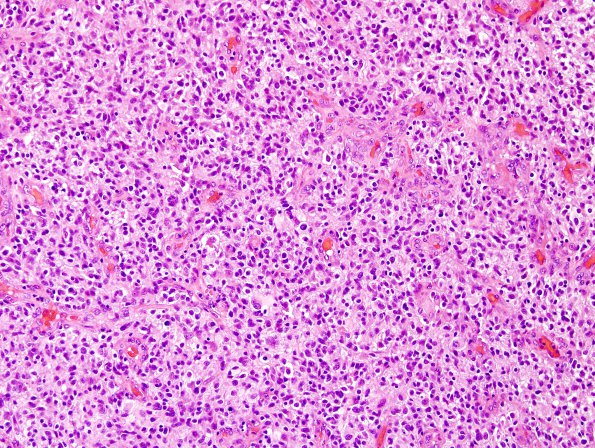
Washington University Experience | NEOPLASMS (GLIAL) | Glioblastoma, small cell type | 5A1 GBM, focal small cell (Case 5) H&E.jpg
Case 5 History ---- The patient was a 52-year-old woman who presented with a seizure. Brain MRI showed a rim-enhancing left parietal lesion. Operative procedure: Stealth-guided left frontal-temporal craniotomy for tumor resection. ---- 5A1-3 Histologic evaluation of the tumor shows a highly cellular proliferation of anaplastic cells with hyperchromatic, round to oval nuclei, prominent nucleoli and moderate amount of cytoplasm. A subset of the cells are round and have clear cytoplasm and well-defined cell borders. Mitotic rate is high. Palisading necrosis is seen at multiple foci.A subset of the cells are round and have clear cytoplasm and well-defined cell borders. Mitotic rate is high. Palisading necrosis and endothelial proliferation are seen at multiple foci.

