Table of Contents
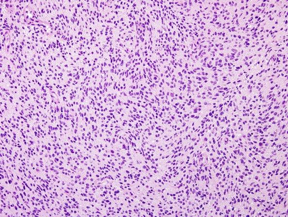
Washington University Experience | NEOPLASMS (GLIAL) | Glioblastoma, small cell type | 7A1 GBM, small cell features (Case 7) H&E 2.jpg
Case 7 History ---- The patient is a 90 year old man with a left frontal lobe neoplasm. ---- 7A1-4 Sections show a hypercellular neoplasm which consists of atypical cells with round and oval nuclei. Some of these have abundant cytoplasm and eccentrically located nuclei characteristic of gemistocytes; whereas, others show a fibrillary appearance. Both features are suggestive of an astrocytic neoplasm. The tumor also shows foci with regular and evenly spaced neoplastic cells with enlarged ovoid round to ovoid nuclei and peri-nuclear cytoplasmic clearing, findings suggestive of oligodendroglial differentiation. Microvascular proliferation and pseudo-palisading necrosis are present. Mitoses are easily identified and number up to 18/10HPF.

