Table of Contents
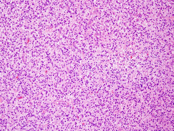
Washington University Experience | NEOPLASMS (GLIAL) | Glioblastoma, small cell type | 8A1 GBM, focal small cell (Case 8) H&E 6
Case 8 History ---- The patient was a 51 year old man who fell following a seizure. Subsequent imaging showed a complex, heterogeneous, right parietal lobe mass with regions of T2 hyperintensity, internal areas of hemorrhage and necrosis, and irregular nodular continuous rim enhancement. Operative procedure: Craniotomy and resection of right parietal tumor. ---- 8A1-3 This hypercellular glial neoplasm consists of densely packed areas of neoplastic cells with round to ovoid nuclei, mildly cleared chromatin with stippling, and inconspicuous nucleoli. They have eosinophilic cytoplasm and poorly defined cell-cell borders. Other areas have more traditional glioblastoma appearance with increased nuclear pleomorphism and small hyperchromatic nuclei with bipolar nuclei. A dense network of vascular channels traverses much of the lesion and there are multiple foci of endothelial hyperplasia present. Mitoses are numerous (17/10HPF). Pseudopalisading necrosis is present in multiple sections of the examined specimen.

