Table of Contents
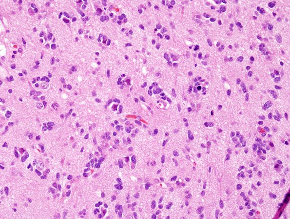
Washington University Experience | NEOPLASMS (GLIAL) | Glioblastoma, small cell type | 9A1 GBM, small cell subtype (Case 9) H&E 9.jpg
Case 9 History ---- The patient was an 18 year old male who presented recently with new onset seizures, headaches, and right sided weakness. A left frontal lobe tumor was identified on CT scan and his symptoms rapidly progressed to an unresponsive state, necessitating surgical intervention. ---- 9A1-3 This is a highly cellular infiltrative glial neoplasm with extensive secondary structure formation, including subpial condensation, perivascular aggregation, and perineuronal satellitosis. There is mild to moderate nuclear pleomorphism, with the majority of tumor nuclei being oval with mild hyperchromasia and minimal discernible cytoplasm. The mitotic index is brisk and there is focal early endothelial hyperplasia. No definite tumor necrosis is seen.

