Table of Contents
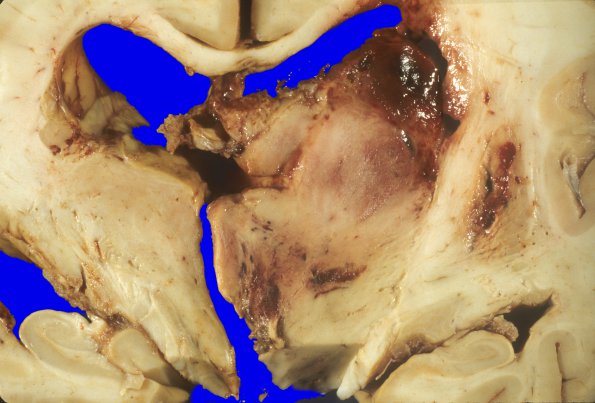
Washington University Experience | NEOPLASMS (GLIAL) | Glioblastoma - Gross Pathology | 10A3 Glioblastoma (Case 31) 3
Higher magnification of image 10A2. ---- Not shown: Sections of the hemorrhagic mass show a neoplasm composed of poorly differentiated astrocytes and small undifferentiated cells with endothelial proliferation, coagulative tumor necrosis with pseudo-palisading of viable tumor cells, numerous mitoses, and superimposed acute hemorrhage. Hemorrhages and edema are demonstrated in the left uncus. Sections of the pons and medulla showed recent Duret hemorrhages.

