Table of Contents
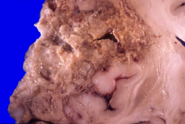
Washington University Experience | NEOPLASMS (GLIAL) | Glioblastoma - Gross Pathology | 11A2 GBM (Case 11) 2
At autopsy a tumor mass was attached to the dura adjacent to the left parietal lobe. There is herniation of the left cingulate gyrus and uncus to the right. Serial coronal sections of the cerebral hemispheres reveal a 6.4 x 7.0 cm, extensively necrotic yellow-tan tumor invading the left frontal, parietal and temporal lobes. The left lateral ventricle is compressed whereas the right is dilated. ---- Not shown: Sections of the tumor show a GBM characterized by sheets of tightly packed, highly pleomorphic glial cells separated by wide regions of tumor necrosis, numerous mitoses and vessels with endothelial proliferation. Microscopic examination of the substantia nigra showed the morphologic alterations associated with Parkinson's disease.

