Table of Contents
Washington University Experience | NEOPLASMS (GLIAL) | Glioblastoma - Gross Pathology | 16A4 Glioblastoma (Case 16) 2
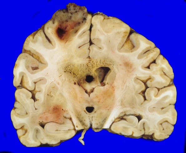
Coronal sections reveal the left frontal lobe and left temporal lobe are two foci that are pink-tan with ill-defined borders with the surrounding tissues. The periventricular areas show a diffuse unusual multicystic appearance. The ventricular system is dilated with a cyst in the septum pellucidum, measuring 3.5 cm in diameter. ---- Not shown: This neoplasm has various alternating histologic appearances that show necrosis and vascular proliferation. Extensive areas of hemorrhage and hyalinization are present and may be secondary to radiation therapy. In addition, vessels with thick, hyalinized walls are seen. The neoplastic cells diffusely infiltrate the brain parenchyma and extend into the midbrain, pons, and medulla. There is also leptomeningeal spread most prominent in the cerebellum.

