Table of Contents
Washington University Experience | NEOPLASMS (GLIAL) | Glioblastoma - Gross Pathology | 18A1 GBM (Case 18) 2
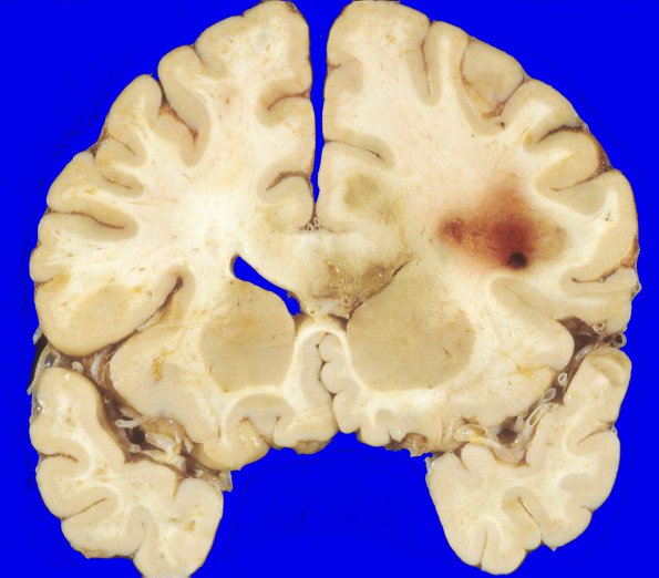
At autopsy, the weight of the unfixed brain is 1180g. Coronal sections of the cerebral hemispheres reveal a right frontal lobe white matter hemorrhagic area measuring 2 cm in greatest dimension, that is soft and irregular. A deeper, yellow-green, irregular, soft and granular lesion is present involving the right cingulate gyrus, right corpus callosum, white matter of the right frontal lobe and angle of the right lateral ventricle. The basal ganglia and thalamus are unremarkable. The ventricular system has an irregular surface in the area corresponding to the deep white matter lesion. ---- Not shown: Sections of the grossly described lesions show a GBM with atypical cells that have hyperchromatic nuclei and abundant eosinophilic cytoplasm, alternating with areas composed of small, bipolar cells. There are extensive areas of necrosis, vascular proliferation, and high mitotic activity. The neoplasm extends to involve the leptomeninges in many sites. The superficial hemorrhagic area on the right frontal lobe surface shows hemorrhage, fibrosis, edema, and histiocytic infiltration, consistent with a previous biopsy site.

