Table of Contents
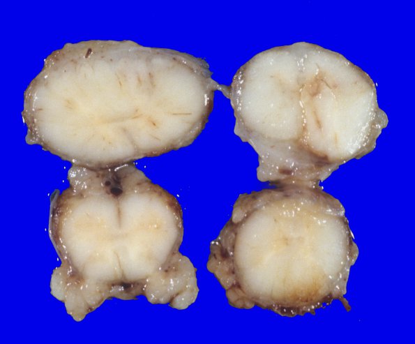
Washington University Experience | NEOPLASMS (GLIAL) | Glioblastoma - Gross Pathology | 19A5 GBM (Case 19) 2
Radial sections of the cerebellum mostly show normal foliar architecture; however, the inferior portion of the vermis is tan, soft and gelatinous consistent with tumor infiltration. The remaining cortex, white matter, and dentate nucleus are unremarkable. ---- Not shown: Multiple sections of the frontal, parietal, temporal and occipital neocortices show disseminated GBM. The tumor is hypercellular and characterized by tumor cells with markedly pleomorphic angulated hyperchromatic nuclei. Mitotic activity is brisk. Both perivascular pseudo-palisading necrosis and microvascular proliferation are evident. Infiltrative tumor cells are present in grey and white matter and some perivascular spaces on all the sections examined, including the ependymal surface of the ventricles. ---- Microscopic examination of the cerebellum shows extensive tumor cell infiltration in the parenchyma of the cerebellar cortex and white matter with focal hemorrhagic necrosis in the central white matter and dentate nucleus. There is extensive tumor infiltrating in the subarachnoid space and, focally, parenchyma of the spinal cord, midbrain, pons and medulla .

