Table of Contents
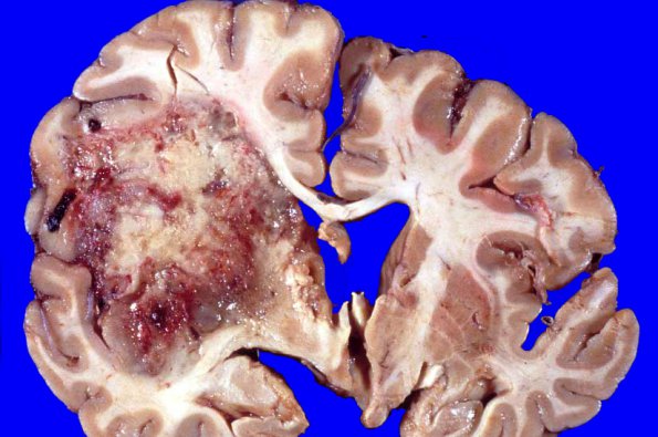
Washington University Experience | NEOPLASMS (GLIAL) | Glioblastoma - Gross Pathology | 1A1 Glioblastoma (Case 1) no Rx 7
1A1,2 Coronal sections showed a large well-circumscribed hemorrhagic mass in the left hemisphere extending from the frontal lobe caudally into the temporal and parietal lobes. In its greatest extent it is 7 cm in diameter and occupies largely the white matter; the cortex overlying the mass is well preserved but shows gyral expansion. Sections of brainstem showed slight atrophy of the left pyramid.

