Table of Contents
Washington University Experience | NEOPLASMS (GLIAL) | Glioblastoma - Gross Pathology | 21A2 GBM (Case 21) 4
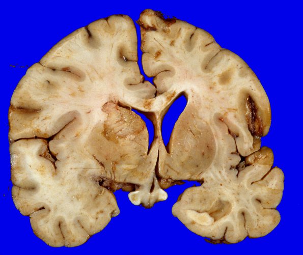
21A2-4 There is subfalcine herniation of the left cingulate gyrus. Coronal sections of the left cerebral hemisphere show a soft pink to yellow, granular lesion in the grey and white matter with expansion of the white matter extending from the left frontal lobe involving the corpus callosum, basal ganglia and parietal cortex, to the occipital lobe. The left lateral ventricle is compressed and displaced to the right. There are foci of superimposed basal ganglia infarcts.

