Table of Contents
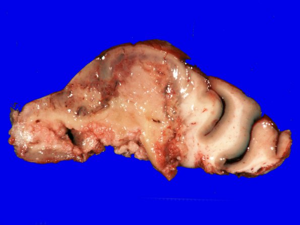
Washington University Experience | NEOPLASMS (GLIAL) | Glioblastoma - Gross Pathology | 24A1 GBM (Case 24) Match with H&E
Case 24 History ---- The patient was a 64 year old man presenting with imbalance, multiple falls and migraines. A CT scan of the brain shows a large hemispheric rim enhancing lesion. Operative procedure: Craniotomy for excision of tumor. ---- 24A1,2 The neurosurgical specimen shows a high-grade glioma with large areas of central necrosis, exuberant microvascular proliferation, hypercellularity, and moderate to marked cytologic atypia. The tumor cells have abundant eosinophilic, fibrillary cytoplasm mostly, enlarged, bipolar to oval nuclei, open to hyperchromatic chromatin and occasional presence of nucleoli. Mitotic figures are easy to find. The edge of the tumor shows an infiltrative growth pattern with perineuronal satellitosis.

