Table of Contents
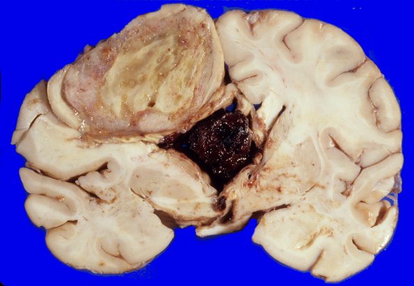
Washington University Experience | NEOPLASMS (GLIAL) | Glioblastoma - Gross Pathology | 25A1 Glioblastoma (Case 25) 1
Coronal sections through the cerebral hemispheres reveal the presence of a large 5 x 4.5 cm mass with a cystic center which extends from the rostral portion of the left frontal pole to the posterior commissure on the left side. The margins of the mass are deceptively well circumscribed, The 3rd ventricle is dilated and filled with a mass of recently clotted blood pushing the fornix to the right. ----
Not shown: Microscopic sections revealed a pleomorphic cellular malignant neoplasm with zones of hemorrhage, necrosis, endothelial capillary proliferation and cystic changes. Pseudopalisading around zones of necrosis, perivascular pseudorosettes and multinucleated giant cells were all features of this neoplasm.

