Table of Contents
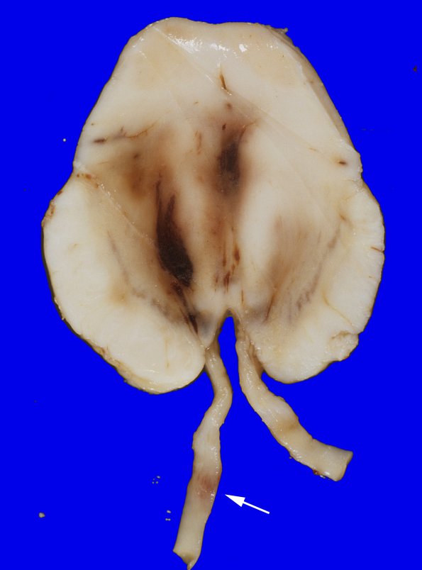
Washington University Experience | NEOPLASMS (GLIAL) | Glioblastoma - Gross Pathology | 27A6 Gliomatosis cerebri, focal GBM (Case 27) 3 copy
The transverse sections of the brainstem show an elongation of the midbrain in the antero-posterior axis and tan-brown, 0.9 x 0.5 x 0.5 cm hemorrhage along the midline (Duret hemorrhages) and also in the left tegmentum. Sections of the pons and medulla show extensive hemorrhage in the tegmentum and reticular formation. Punctate hemorrhage is also noted in the basis pontis. Also noted is a 0.5 x 0.5 cm area of tan, brown discoloration in the left third nerve (arrow, 27A6). These changes are consistent with uncal herniation. ---- Not shown: The left temporal hemorrhagic area is composed of a discrete high grade mass predominantly composed of gemistocytic cells. Areas of pseudopalisading necrosis and endothelial hyperplasia are both identified. These findings are consistent with an astrocytic gliomatosis with focal transformation to glioblastoma multiforme. Neoplasm is seen within the left cerebral peduncle of midbrain, descending tracts in basis pontis and left pyramid in the medulla. The pons and the midbrain have multiple foci of hemorrhagic infarcts. In addition, the patient also demonstrated extensive infarcts and hemorrhagic infarcts in the neocortices and other regions. These changes may be secondary to the combination of several factors, such as expansion of the brain parenchyma by tumor infiltration, intracranial bleeding and uncal herniation, which result in significant decrease in blood circulation into brain.

