Table of Contents
Washington University Experience | NEOPLASMS (GLIAL) | Glioblastoma - Gross Pathology | 2A2 GBM (Case 2) 9
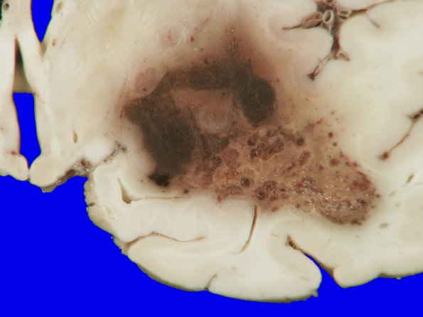
Coronal sections of the cerebral hemispheres show a tan-red, hemorrhagic and centrally necrotic lesion present in the right temporal lobe, measuring 3 x 2.5 x 2 cm, which expands the adjacent temporal lobe compared to the contralateral side. ---- Not shown: This is a high grade glial neoplasm composed of cells with oval to elongate nuclei and scant cytoplasm as well as focal areas with a gemistocytic appearance, numerous mitotic figures, microvascular proliferation, pseudopalisading necrosis, fibrinoid vascular necrosis and hemorrhage. Sections of the right basal ganglia, right thalamus, right hippocampus and right middle cerebral artery show infiltrating glial neoplasm with perivascular arrangement of tumor cells and extension into the subarachnoid space as well as reentry of the tumor via the Virchow-Robin spaces.

